LABORATORIES
Cajal Blue Brain Platform
Cajal Blue Brain Platform CSIC/UPM
Research
Team
Publications
Contact
Others
Research
One of the main goals of neuroscience is to understand the biological mechanisms responsible for human mental activity. In particular, the study of the cerebral cortex is and without any doubt will be the greatest challenge for science in the next centuries, since it represents the foundation of our humanity. In other words, the cerebral cortex is the structure whose activity is related to the capabilities that distinguish humans from other mammals. Thanks to the development and evolution of the cerebral cortex we are able to perform highly complex and specifically human tasks, such as writing a book, composing a symphony or developing technologies. For these reasons, the study of the normal brain is obviously necessary to better understand not only how our brain functions under normal conditions, but also how its alterations give rise to many devastating brain diseases like AD. Indeed, the Cajal Blue Brain node (CSIC/UPM) was created as a basic research unit located at the Montegancedo Campus (UPM, Madrid), as a continuation of the Cajal Blue Brain Project (https://cajalbbp.es/). The node is aimed to study the structural and functional organization of the human brain (both in health and disease) as well as the brain of different experimental animals, coordinating different levels of analysis (molecules, synapses, neurons, local circuits, brain networks function and modelling). Also, a major objective of the node is to develop new techniques and strategies for studying brain structural organization and function.
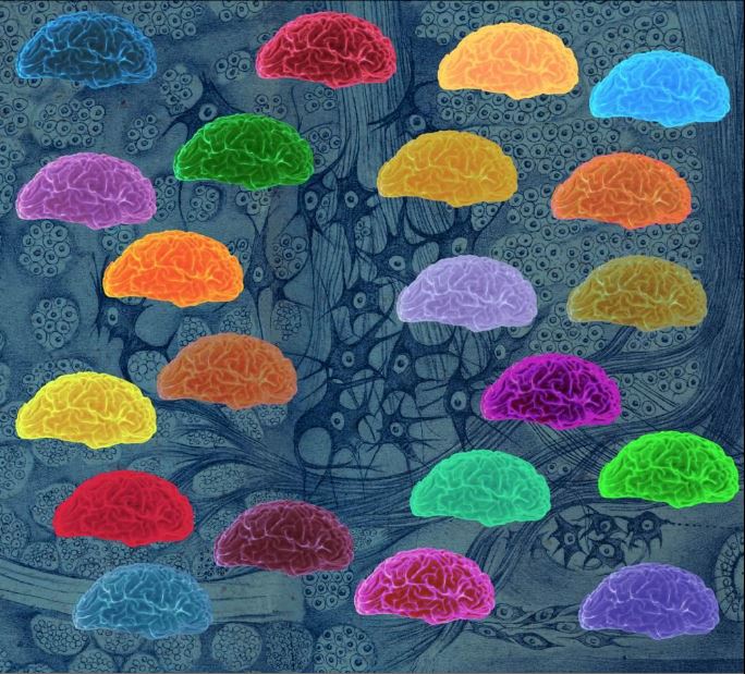
In 2009, the Cajal Blue Brain project concession (https://cajalbbp.csic.es/) launched a new stage for the laboratory’s scientific work. This project allowed the creation of a multidisciplinary team with more than 50 researchers (neuroanatomists, physiologists, mathematicians and computer engineers) and the establishment of the Cajal Laboratory of Cortical Circuits (LCCC) under a UPM/CSIC agreement. Thanks to this, it has been possible to develop new anatomical and computational tools and methods that have made an important technological contribution to the study of the brain. One of these tools is three-dimensional electron microscopy (FIB/SEM) technology, which enables three-dimensional reconstruction of brain tissue at the ultrastructural level. This novel technology has proven to be essential for deciphering the synaptic map or synaptome.
Lines of investigation
1. High throughput brain-wide screening of morphological and neurochemical types of synapses and cells
2. Detailed single cell studies using cell filling
3. Detailed synapse studies using 3D electron microscopy
4. Informatics and modelling
Team

Dr. Javier DeFelipe (CSIC)
Research Proffessor

Ruth Benavides Piccione (CSIC)
Tenured scientist
José Rodrigo Rodríguez Sánchez (CSIC)
Tenured scientist
Lidia Alonso Nanclares (CSIC)
Scientific Researcher
Ángel Merchán Pérez (UPM)
Scientific Researcher
Paula Merino Serrais (CSIC)
Scientific Researcher

Marta Turegano (CSIC)
Scientific Researcher
Lidia Blázquez Llorca (UCM)
Scientific Researcher
Nicolás Cano Astorga (CSIC)
Predoctoral Researcher
Sergio Plaza Alonso (CSIC)
Predoctoral Researcher

Isabel Fernaud Espinosa (CSIC)
Technician
Ana Isabel García Ramírez (CSIC)
Technician

Asta Kastanaukaiste (CSIC)
Technician
Alberto Muñoz Céspedes (UCM) Scientific Researcher
Gonzalo León Espinosa (CEU) Scientific Researcher
Lorena Valdés Lora (CSIC) Technician
Pilar Flores Romero (UPM) Project Manager
Publications
Publications of the last 5 years
- León-Espinosa G, Regalado-Reyes M, DeFelipe J, Muñoz A (2018) Changes in neocortical and hippocampal microglial cells during hibernation. Brain Struct Funct. 223:1881–1895. https://doi.org/10.1007/s00429-017-1596-7
- Santuy A, Rodriguez JR, DeFelipe J, Merchan-Perez A (2018) Volume electron microscopy of the distribution of synapses in the neuropil of the juvenile rat somatosensory cortex. Brain Struct Funct. 223:77-90. https://doi.org/10.1007/s00429-017-1470-7
- Santuy A, Rodríguez JR, DeFelipe J, Merchán-Pérez A (2018) Study of the size and shape of synapses in the juvenile rat somatosensory cortex with 3D Electron Microscopy. eNeuro 5(1), ENEURO.0377-17.2017. https://doi.org/10.1523/eneuro.0377-17.2017
- Domínguez-Álvaro M,Montero-Crespo M, Blázquez-Llorca L, Insausti R, DeFelipe J, AlonsoNanclares L (2018) Three-dimensional analysis of synapses in the transentorhinal cortex of Alzheimer’s disease patients. Acta Neuropathol Commun. 6:20. https://doi.org/10.1186/s40478-018-0520-6
- Rodríguez J-R, Turégano-López M, DeFelipe J and Merchán-Pérez A (2018) Neuroanatomy from mesoscopic to nanoscopic scales: An improved method for the observation of semithin sections by high-resolution scanning electron microscopy. Front. Neuroanat. 12:14. https://doi.org/10.3389/fnana.2018.00014
- Varando G, Benavides-Piccione R, Muñoz A, Kastanauskaite A, Bielza C, Larrañaga P, DeFelipe J (2018) MultiMap: A tool to automatically extract and analyse spatial microscopic data from large stacks of confocal microscopy images. Front. Neuroanat. 12:37. https://doi.org/10.3389/fnana.2018.00037
- Luengo-Sanchez S, Fernaud-Espinosa I, Bielza C, Benavides-Piccione R, Larrañaga P, DeFelipe J (2018) 3D morphology-based clustering and simulation of human pyramidal cell dendritic spines. PLoS Comput Biol. 14(6):e1006221. https://doi.org/10.1371/journal.pcbi.1006221
- Furcila D, DeFelipe J, Alonso-Nanclares L (2018) A Study of Amyloid-β and Phosphotau in Plaques and Neurons in the Hippocampus of Alzheimer’s Disease Patients. J Alzheimers Dis. 64:417-435. https://doi.org/10.3233/jad-180173
- Fernández-Cabrera MR, Higuera-Matas A, Fernaud-Espinosa I, DeFelipe J, Ambrosio E, Miguéns M (2018)Selective effects of Δ9-tetrahydrocannabinol on medium spiny neurons inthe striatum. PLoS One. https://doi.org/10.1371/journal.pone.0200950
- Eyal G, Verhoog MB, Testa-Silva G, Deitcher Y, Benavides-Piccione R, DeFelipe J, de Kock CPJ, Mansvelder HD, Segev I (2018) Human cortical pyramidal neurons: from spines to spikes via models. Front Cell Neurosci. 12:181. https://doi.org/10.3389/fncel.2018.00181
- Roy M, Sorokina O, McLean C, Tapia-González S, DeFelipe J, Armstrong JD, Grant SGN (2018) Regional diversity in the postsynaptic proteome of the mouse brain. Proteomes. https://doi.org/10.3390/proteomes6030031
- Santuy A, Turégano-López M1, Rodríguez JR, Alonso-Nanclares L, DeFelipe J, Merchán-Pérez A (2018) A Quantitative study on the distribution of mitochondria in the neuropil of the juvenile rat somatosensory cortex. Cereb Cortex 28:3673–3684. http://dx.doi.org/10.1093/cercor/bhy159
- Zhu F, Cizeron M, Qiu Z, Benavides-Piccione R, Kopanitsa MV, Skene NG, Koniaris B, DeFelipe J, Fransén E, Komiyama NH3, Grant SGN (2018) Architecture of the mouse brain synaptome. Neuron 99:781-799. https://doi.org/10.1016/j.neuron.2018.07.007
- León-Espinosa G, Antón-Fernández A, Tapia-González S, DeFelipe J, Muñoz A (2018) Modifications of the axon initial segment during the hibernation of the Syrian hamster. Brain Struct Funct. 223:4307-4321. https://doi.org/10.1007/s00429-018-1753-7
- Rockland KS and DeFelipe J (2018) Editorial: Why Have Cortical Layers? What Is the function of layering? Do neurons in cortex integrate information across different layers? Front Neuroanat. 12:78. https://doi.org/10.3389/fnana.2018.00078
- Merino-Serrais P, Loera-Valencia R, Rodriguez-Rodriguez P, Parrado-Fernandez C, Ismail MA, Maioli S, Matute E, Jimenez-Mateos EM, Björkhem I, DeFelipe J, Cedazo-Minguez A (2018) 27- Hydroxycholesterol induces aberrant morphology and synaptic dysfunction in hippocampal neurons. Cereb Cortex 29:429-446. https://doi.org/10.1093/cercor/bhy274
- Mihaljević B,Larrañaga, P, Benavides-Piccione R, Hill S, DeFelipe J, & Bielza C (2018) Towards a supervised classification of neocortical interneuron morphologies. BMC bioinformatics, 19(1), 511. https://doi.org/10.1186/s12859-018-2470-1
- Aliaga Maraver JJ, Mata S, Benavides-Piccione R, DeFelipe, J, Pastor L (2018) A Method for the symbolic representation of neurons. Front Neuroanat. 12:106. https://doi.org/10.3389/fnana.2018.00106
- Kwon T, Merchán-Pérez A, Rial Verde EM, Rodríguez JR, DeFelipe J, Yuste R (2019) Ultrastructural, molecular and functional mapping of GABAergic synapses on dendritic spines and shafts of neocortical pyramidal neurons. Cereb Cortex 29:2771–2781. https://doi.org/10.1093/cercor/bhy143
- Gonzalez-Riano C, Leon-Espinosa G, Regalado-Reyes M, García A, DeFelipe J, Barbas C (2019) A Metabolomic study of hibernating Syrian hamster brain: in search of neuroprotective agents J Proteome Res. 18:1175–1190 https://doi.org/10.1021/acs.jproteome.8b00816
- León-Espinosa G, DeFelipe J, Muñoz A (2019) The Golgi apparatus of neocortical glial cells during hibernation in the Syrian hamster. Front Neuroant.13: 92. https://doi.org/10.3389/fnana.2019.00092
- Leguey I, Benavides-Piccione R, Rojo C, Larrañaga P, Bielza C, DeFelipe J (2019) Patterns of dendritic basal field orientation of pyramidal neurons in the rat somatosensory cortex. eNeuro, 5(6), ENEURO.0142-18.2018. https://doi.org/10.1523/eneuro.0142-18.2018
- Regalado-Reyes M, Furcila D, Hernández F, Ávila J, DeFelipe J, León-Espinosa G (2019) Phospho-tau changes in the human CA1 during Alzheimer’s disease progression. J Alzheimers Dis, 69(1): 277–288. https://doi.org/10.3233/JAD-181263
- Domínguez-Álvaro M, Montero-Crespo M, Blázquez-Llorca L, DeFelipe J, Alonso-Nanclares L (2019) 3D electron microscopy study of synaptic organization of the normal human transentorhinal cortex and its possible alterations in Alzheimer’s disease. eNeuro. pii: ENEURO.0140-19.2019 https://doi.org/10.1523/eneuro.0140-19.2019
- Furcila D, Domínguez-Álvaro M, DeFelipe J, Alonso-Nanclares L (2019) Subregional density of neurons, neurofibrillary tangles and amyloid plaques in the hippocampus of patients with Alzheimer’s disease. Front Neuroanat. 13:99. https://doi.org/10.3389/fnana.2019.00099
- Ortuño T, López-Madrona VJ, Makarova J, Tapia-González S, Muñoz A, DeFelipe J, Herreras O. 019. Slow-wave activity in the S1hl cortex is contributed by different layer-specific field potential sources during development. J Neurosci. 39:8900–8915. https://doi.org/10.1523/jneurosci.1212-19.2019
- Mihaljević B, Benavides-Piccione R, Bielza C, Larrañaga P, DeFelipe J (2019) Classification of GABAergic interneurons by leading neuroscientists. Sci Data. 26(1):221. https://doi.org/10.1038/s41597-019-0246-8
- Tapia-González S, Insausti R, DeFelipe J (2019) Differential expression of secretagogin immunostaining in the hippocampal formation and the entorhinal and perirhinal cortices of humans, rats, and mice. J Comp Neurol 528:523-541. https://doi.org/10.1002/cne.24773
- Benavides-Piccione R, Regalado-Reyes M, Fernaud-Espinosa I, Kastanauskaite A, TapiaGonzález S, León-Espinosa G, Rojo C, Insausti R, Segev I, DeFelipe J (2020) Differential structure of hippocampal CA1 pyramidal neurons in the human and mouse. Cereb Cortex 30:730- 752. https://doi.org/10.1093/cercor/bhz122
- Turegano-Lopez M, Santuy A, DeFelipe J, Merchan-Perez A (2020) Size, shape, and distribution of multivesicular bodies in the juvenile rat somatosensory cortex: A 3D electron microscopy study. Cereb Cortex. 30:1887-1901. http://dx.doi.org/10.1093/cercor/bhz211
- Merino-Serrais P, Tapia-González S, DeFelipe J (2020) Calbindin immunostaining in the CA1 hippocampal pyramidal cell layer of the human and mouse: A comparative study. J Chem Neuroanat. 104:101745. https://doi.org/10.1016/j.jchemneu.2020.101745
- Kikuchi T, Gonzalez-Soriano J, Kastanauskaite A, Benavides-Piccione R, Merchan-Perez A, DeFelipe J, Blazquez-Llorca L (2020) Volume electron microscopy study of the relationship between synapses and astrocytes in the developing rat somatosensory cortex. Cereb Cortex. 30:3800–3819. https://doi.org/10.1093/cercor/bhz343
- Montero-Crespo M, Domínguez-Álvaro M, Rondón-Carrillo P, Alonso-Nanclares L, DeFelipe J, Blázquez-Llorca L (2020) Three-dimensional synaptic organization of the human hippocampal CA1 field. eLife 2020;9:e57013. https://doi.org/10.7554/eLife.57013
- Rodriguez-Moreno J, Porrero C, Rollenhagen A, Rubio-Teves M, et al (2020) Area-specific synapse structure in branched posterior nucleus axons reveals a new level of complexity in thalamocortical networks. J Neurosci. 40:2663 2679. https://doi.org/10.1523/JNEUROSCI.2886-19.2020
- Yuste R, Hawrylycz M, Aalling N, Aguilar-Valles A, Arendt D, et al (2020) A community-based transcriptomics classification and nomenclature of neocortical cell types. Nat Neurosci. 23:1456- 1468. https://dx.doi.org/10.1038%2Fs41593-020-0685-8
- Mihaljević B, Larrañaga P, Benavides-Piccione R, DeFelipe J, Bielza C (2020) Comparing basal dendrite branches in human and mouse hippocampal CA1 pyramidal neurons with Bayesian networks. Sci Rep. 10(1):18592. https://doi.org/10.1038/s41598-020-73617-9
- Santuy A, Tomás-Roca L, Rodríguez JR, González-Soriano J, Zhu F, Qiu Z, Grant SGN, DeFelipe J, Merchan-Perez A (2020) Estimation of the number of synapses in the hippocampus and brainwide by volume electron microscopy and genetic labeling. Sci Rep. 10(1):14014. https://doi.org/10.1038/s41598-020-70859-5
- Velasco I, Toharia P, Benavides-Piccione R, Fernaud-Espinosa I, Brito JP, Mata S, DeFelipe J, Pastor L, Bayona S (2020) Neuronize v2: bridging the gap between existing proprietary tools to optimize neuroscientific workflows. Front Neuroanat. 14:585793. https://doi.org/10.3389/fnana.2020.585793
- Regalado-Reyes M, Benavides-Piccione R, Fernaud-Espinosa I, DeFelipe J, León-Espinosa G (2020) Effect of phosphorylated tau on cortical pyramidal neuron morphology during hibernation. Cereb Cortex Communications. 1:1-25. https://doi.org/10.1093/texcom/tgaa018
- Domínguez-Álvaro M, Montero-Crespo L Blazquez-Llorca, DeFelipe J, Alonso-Nanclares L (2021) 3D Ultrastructural study of synapses in the human entorhinal cortex. Cereb Cortex. 31:410–425. https://doi.org/10.1093/cercor/bhaa233
- DeFelipe J, De Carlos JA, Metha AR (2021) A museum for Cajal’s Legacy. The Lancet Neurology.20(1):25. https://doi.org/10.1016/S1474-4422(20)30444-0
- Montero-Crespo M, Domínguez-Álvaro M, Alonso-Nanclares L, DeFelipe J, Blazquez-Llorca L (2021) Three-dimensional analysis of synaptic organization in the hippocampal CA1 region in Alzheimer’s disease. Brain. 144(2):553-573. https://doi.org/10.1093/brain/awaa406
- Loera-Valencia R, Vazquez-Juarez E, Muñoz A, Gerenu G, Gómez-Galán M, Lindskog M, DeFelipe J, Cedazo-Minguez A, Merino-Serrais P (2021) High levels of 27-hydroxycholesterol results in synaptic plasticity alterations in the hippocampus. Sci Rep. 11(1):3736. https://doi.org/10.1038/s41598-021-83008-3
- Blazquez-Llorca L, Miguéns M, Montero-Crespo M, Selvas A, Gonzalez-Soriano J, Ambrosio E, DeFelipe J (2021) 3D synaptic organization of the rat CA1 and alterations induced by cocaine self-administration. Cereb Cortex. 31(4):1927-1952. https://doi.org/10.1093/cercor/bhaa331
- Gonzalez-Riano C, Tapia-González S, Perea G, González-Arias C, DeFelipe J, Barbas C (2021)Metabolic changes in brain slices over time: a multiplatform metabolomics approach. Mol Neurobiol. 58:3224–3237. https://doi.org/10.1007/s12035-020-02264-y32
- Benavides-Piccione R, Rojo C, Kastanauskaite A, DeFelipe J (2021) Variation in pyramidal cellmorphology across the human anterior temporal lobe. Cereb cortex 31(8):3592–3609. https://doi.org/10.1093/cercor/bhab034
- Cano-Astorga N, DeFelipe J, Alonso-Nanclares L(2021) Three-dimensional synaptic organization of layer III of the human temporal neocortex. Cereb Cortex bhab120. https://doi.org/10.1093/cercor/bhab120
- Maestú F, de Haan W, Busche MA, DeFelipe J. 2021. Neuronal excitation/inhibition imbalance: core element of a translational perspective on Alzheimer pathophysiology. Ageing Research Reviews. 69:101372. https://doi.org/10.1016/j.arr.2021.101372
- Domínguez-Álvaro M, Montero-Crespo M, Blazquez-Llorca L, Plaza-Alonso S, Cano-Astorga N, DeFelipe J, Alonso-Nanclares L (2021) 3D analysis of the synaptic organization in the entorhinal cortex in Alzheimer’s disease. eNeuro.0504-20.2021. https://doi.org/10.1523/ENEURO.0504-20
- LaTorre A, Alonso-Nanclares L, Peña JM, DeFelipe J (2021) 3D segmentation of neuronal nuclei and cell-type identification using multi-channel information. Expert Syst Appl. 183:115443. https://doi.org/10.1016/j.eswa.2021.115443
- Luján R, Merchán-Pérez A, Soriano J, Martín-Belmonte A, Aguado C, Alfaro-Ruiz R, MorenoMartínez AE and DeFelipe J (2021) Neuron class and target variability in the three-dimensional localization of sk2 channels in hippocampal neurons as detected by immunogold FIB-SEM. Front. Neuroanat. 15:781314. doi: 10.3389/fnana.2021.781314
- Vidaurre-Gallart I, Fernaud-Espinosa I, Cosmin-Toader N, Talavera-Martínez L, Martin-Abadal M, Benavides-Piccione R, Gonzalez-Cid Y, Pastor L, DeFelipe J and García-Lorenzo M (2022) A deep learning-based workflow for dendritic spine segmentation. Front. Neuroanat. 16:817903. doi: 10.3389/fnana.2022.817903
- Amunts K, DeFelipe J, Pennartz C, Destexhe A, Migliore M, Ryvlin P, Furber S, Knoll A, Bitsch L, Bjaalie JG, Ioannidis Y, Lippert T, Sanchez-Vives MV, Goebel R, Jirsa V (2022). Linking brain structure, activity and cognitive function through computation. eNeuro. ENEURO.0316- 21.2022. doi: 10.1523/ENEURO.0316-21.2022
- Merino-Serrais P, Plaza-Alonso S, Hellal F, Valero-Freitag S, Kastanauskaite A, Muñoz A, Plesnila N, DeFelipe J (2022). Microanatomical study of pyramidal neurons in the contralesional somatosensory cortex after experimental ischemic stroke. Cereb Cortex bhac121. doi: 10.1093/cercor/bhac121.
- Turegano-Lopez M, Santuy A, Kastanauskaite A, Rodriguez JR, DeFelipe J, Merchan-Perez A (2022). Single-neuron labeling in fixed tissue and targeted volume electron microscopy. Front Neuroanat. 16:852057. doi: 10.3389/fnana.2022.852057.
- Antón-Fernández A, León-Espinosa,G, DeFelipe J, Muñoz A (2022). Pyramidal cell axon initial segment in Alzheimer´s disease. Sci Rep 12, 8722. https://doi.org/10.1038/s41598-022-12700-9
- Chindemi G, Abdellah M, Amsalem O, Benavides-Piccione R, Delattre V, Doron M, Ecker A, King J, Kumbhar P, Monney C, Perin R, Rössert C, Van Geit W, DeFelipe J, Graupner M, Segev I, Markram H, Muller E. (2022). A calcium-based plasticity model predicts long-term potentiation and depression in the neocortex. Nat Commun 13, 3038. https://doi.org/10.1038/s41467-022-30214-w
- Hunt S, Leibner Y, Mertens EJ, Barros-Zulaica N, Kanari L, Heistek TS, Karnani MM, Aardse R,Wilbers R, Heyer DB, Goriounova NA, Verhoog MB, Testa-Silva G, Obermayer J, Versluis T, Benavides-Piccione R, de Witt-Hamer P, Idema S, Noske DP, Baayen JC, Lein ES, DeFelipe J, Markram H, Mansvelder HD, Schürmann F, Segev I, de Kock CPJ (2022). Strong and reliable 33 synaptic communication between pyramidal neurons in adult human cerebral cortex. Cereb Cortex. bhac246. https://doi: 10.1093/cercor/bhac246
- DeFelipe J (2022) Manifesto of a neuroanatomist. Front. Neuroanat. 16:931547. https://www.frontiersin.org/articles/10.3389/fnana.2022.931547/full
- Bulovaite E, Qiu Z, Kratschke M, Zgraj A, Fricker DG, Tuck EJ, Gokhale R, Koniaris B, Jami SA, Merino-Serrais P, Husi E, Mendive-Tapia L, Vendrell M, O’Dell TJ, DeFelipe J, Komiyama NH, Holtmaat A, Fransén E, Grant SGN. A brain atlas of synapse protein lifetime across the mouse lifespan. Neuron. 2022 Sep 28:S0896-6273(22)00814-5. doi: 10.1016/j.neuron.2022.09.009.
- Ostos, S, Aparicio, G, Fernaud-Espinosa, I, DeFelipe, J and Muñoz A (2022) Quantitative analysis of the GABAergic innervation of the soma and axon initial segment of pyramidal cells in the human and mouse neocortex. Cereb Cortex. bhac314. doi: 10.1093/cercor/bhac314
- Alonso-Nanclares, L., Rodríguez, J. R., Merchan-Perez, A., González-Soriano, J., Plaza-Alonso, S., Cano-Astorga, N., et al. Cortical synapses of the world’s smallest mammal: An FIB/SEM study in the Etruscan shrew. J Comp Neurol. doi: 10.1002/cne.25432
- DeFelipe J, DeFelipe-Oroquieta J, Furcila D, Muñoz-Alegre M, Maestú F, Sola RG, BlázquezLlorca L, Armañanzas R, Kastanaskaute A, Alonso-Nanclares L, Rockland KS and Arellano JI (2022) Neuroanatomical and psychological considerations in temporal lobe epilepsy. Front. Neuroanat. 16:995286. doi: 10.3389/fnana.2022.995286
Contact
Where to find us
Cajal Laboratory of Cortical Circuits
Centro de Tecnología Biomédica (CTB), Universidad Politécnica de Madrid, Campus Montegancedo S/N Pozuelo de Alarcón 28223 Madrid Spain
Instituto Cajal (CSIC) Avenida Doctor Arce 37 28002 Madrid
Call us
Phone:
Write us
Email address:
Others
Reources
The platform (600 m2) is located in the UPM-Montegancedo Campus and has dedicated bench space for 15 to 20 investigators.
Laboratory, confocal and electron microscopy facilities:
A large amount of the space is dedicated to laboratory equipment. This includes reagents and devices for various histological and immunohistochemical techniques, a Leicavibratome and a cryostate (Thermo) for sectioning tissue, as well as a separate room housing 4 microscope set-ups for intracellular injections in fixed tissue. In addition, the laboratory has an area dedicated to small animal perfusion, and a fully equipped electron microscopy laboratory with a rotary microtome (Microm), laboratory microwave oven (Ted Pella), a pyramidotome (Leica EM Trim) for tissue preparation and an Ultracut (Leica EM UC6) to obtain semithin and ultrathin sections. It includes also an Image Analysis room with 7 computers for image analysis (Imaris software), two microscopes connected to computers with software for image acquisition and analysis (Neurolucida and Stereo Investigator) and the Confocal Microscopy Facility.
The Confocal Microscopy Facility, includes two laser scanning confocal microscopes: a Zeiss LSM 710 with 5 laser-heads (405, 488, 543, 594 and 633nm) and a Leica SP8 AOBS, WLL Laser + Argon (458, 476, 488, 496, 514,) and 405 lasers and 2PMT+2HyD. This facility also has two direct microscopes with motorized stages connected to two computer workstations and two additional workstations capable of image processing and deconvolution, 3-D reconstruction, etc.
It also has a separate room housing a dual-beam electron microscope (Crossbeam 540 electron microscope, Carl Zeiss NTS GmbH, Oberkochen, Germany) and its auxiliary equipment, which is capable of acquiring serial high-resolution images of neural tissue. Additionally, Dr. DeFelipe has a transmission electron microscope (Jeol 1011, 100 Kv; equipped with an 11 MpxGatan-Orius camera), and four computer workstations for electron microscope imaging processing, 3-D reconstruction and analysis — using software (e.g., ESPINA) developed for the Cajal Blue Brain Project (https://cajalbbp.es/). Other equipment includes an icemaker, a Millipore water purification system, a glassware washer, 3 chemical fume hoods, water baths, microscope slide warmers, refrigerators, and other small pieces of laboratory equipment.
Most important projects las 10 years
1. Title: Human Brain Project (FET Flagship SGA2)
Funding agency: EC.
Duration: 01/04/2018-31/03/2020.
Leader Subproject 1: Javier DeFelipe
Amount: 1.280.292€
2. Title: The Pyramidal Neuron in Cognition and Alzheimer’s Disease
Funding agency: Alzheimer’s Association (2014).
Duration: 01/02/2015-31/07/2018.
Principal Investigator: Javier DeFelipe (Instituto Cajal)
Amount: 450.000$
3. Title: Estudio de la microorganización de la corteza cerebral en pacientes de Alzheimer y del hámster como modelo para estudiar la fosforilación de Tau
Funding agency: Ministerio de Economía y Competitividad. Ref.: SAF2015-66603-P
Duration: 2016-2018.
Principal investigator: Javier DeFelipe
Amount: 237.160€
4. Title: Human Brain Project (FET Flagship SGA1)
Funding agency: EC.
Duration: 01/04/2016-31/03/2018.
Co-Leader Subproject 1: Javier DeFelipe
Amount: 1.322.944€
5. Títle: Cajal Blue Brain
Funding agency: Ministerio de Ciencia e Innovación. / Blue Brain/ École Polytechnique fédérale de Lausanne.
Duration: 01/01/2009-31/12/2018.
Director: Javier DeFelipe
Amount: 25M € loan in deposit
6.Title: Microanatomical and Neurochemical Alterations of the cerebral cortex in Alzheimer’s disease.
Funding agency: Ministerio de Economía y Competitividad. BFU2012-34963
Duratio: 01/01/2013-31/12/2015
Principal Investigator: J. DeFelipe
Amount: 193.050 €
7. Title: Estudio microanatómico de la corteza cerebral en pacientes con enfermedad de alzheimer y en modelos animales. Efecto de los cannabinoides en la progresión de la enfermedad.
Funding agency: Ministerio de Ciencia e Innovación. SAF2009-09394 (subprograma NEF).
Duration: 01/01/2010-31/12/2012.
Principal Investigator: J. DeFelipe
Amount: 145.200€

Neuroscience Research Center dependent on the CSIC. Founded in 1920 and initially directed by Santiago Ramón y Cajal. World reference in the study of the brain. Custodian of the Cajal Legacy.
Activities
