WILD CARD OF SWORDS
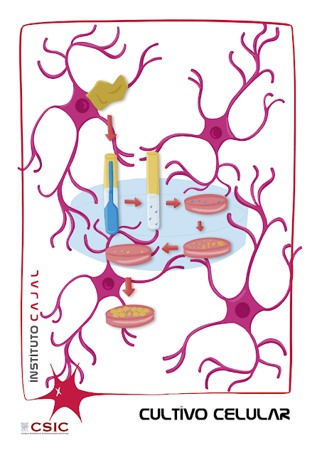
CELL CULTURE
Wild Card of Swords: CELL CULTURE, a very current century-old technique, halfway between science and art

The first CELL CULTURE techniques were developed more than 100 years ago and, since then, have allowed enormous advances in science. Today it is a fundamental tool used in laboratories around the world to study the physiology and biochemistry of cells, the mechanisms underlying disease, and the effects of drugs and toxic compounds on cell populations.
CELL CULTURE is an in vitro study model that has become a basic research technique in laboratories such as the Cajal Institute. It involves growing and proliferating cells—eukaryotic or prokaryotic—in artificially controlled environments with specific nutrients. We are talking about cultures of plant cells, animals, bacteria, yeasts and, lately, also embryonic stem cells.
Cell culture requires a set of techniques that allow the maximum preservation of the physiological, biochemical and genetic properties of those cells that are intended to be maintained in vitro. For this reason, the cell culture laboratory is a place where conditions of total environmental asepsis must be maintained to avoid contamination of the cultures. Since 1987, the Cell Culture Unit of the Cajal Institute is made up of three rooms where cells obtained from multicellular organisms are generated in vitro, especially neurons, astrocytes and oligodendrocytes extracted from animal brains.
According to current law [1], the management of these crops requires two different levels of security (or containment levels):
– Containment level 1: in the Crop Room and Established Lines. Required for culturing non-transformed animal cells from non-primates. It consists of 4 horizontal laminar flow cabins for preferential protection of the samples and another 4 vertical laminar flow cabins that provide protection to the operator as well as the sample.
– Containment level 2: in Rooms P2-A and P2-B, where primate cell cultures (tumor or non-tumor), non-intervened human cell lines, virus-producing non-primate cell cultures are generated, biological agents are manipulated. non-infectious through the air with human tropism (retrovirus, lentivirus, adenovirus). The Cajal Institute is equipped with two biosafety cabins that guarantee protection to the operator, while also providing protection to the sample.
The basic equipment of a cell culture laboratory is as follows:
In addition to having the appropriate equipment and properly aseptic spaces, it is essential to choose an appropriate culture medium. Along with contamination problems, culture media are the main factors involved in the greater or lesser success of in vitro cell culture. Each type of cell culture requires a specific medium, without there being a universal and precise formula for it.
A good growing medium is one that has the following qualities:
– it is specific with respect to the cell to be cultured;
– provides essential nutrients (carbohydrates, essential fatty acids and other lipids, amino acids, mineral salts and trace elements;
– has an adequate pH value;
– has buffer capacity (to maintain the pH within physiological values);
– it is isotonic and, of course,
– it is sterile.
Nowadays, numerous companies market a series of standard base culture media [2] and other more specific ones that are completed with other additives adapted to the type of culture being carried out, such as antibiotics, pH indicators, or even fetal serum. (sheep, bovine or human).
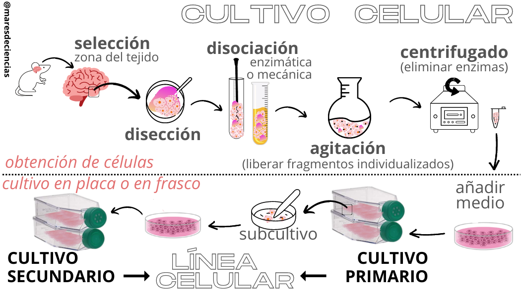
By combining good laboratory practices with the appropriate equipment and materials, it is possible to obtain well-defined clones of a single type of cell from each culture, called cell lines. This is a diagram of the process of obtaining the cells that will be used to obtain them:

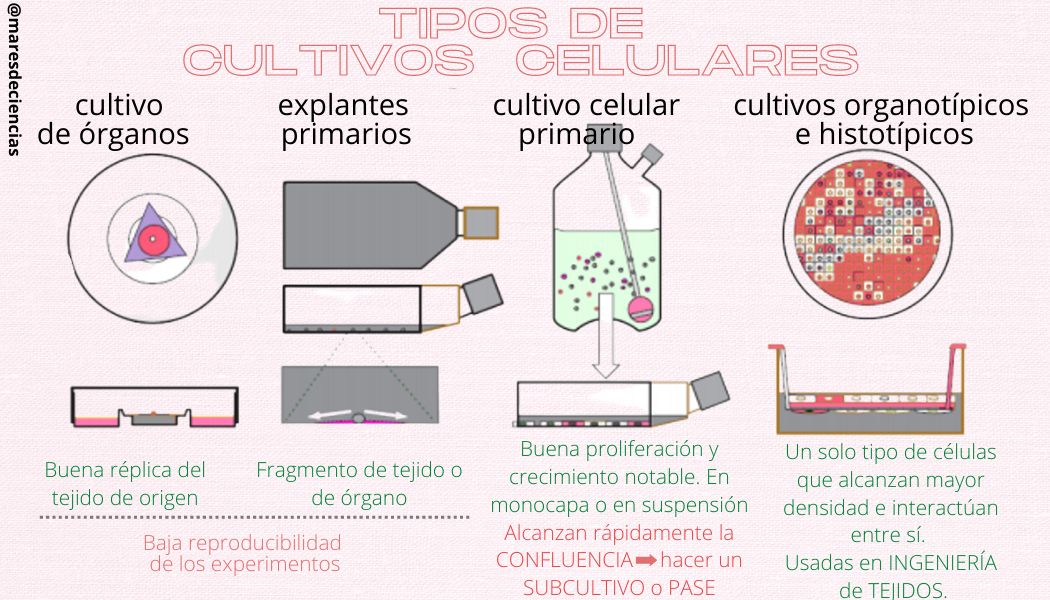
There are several types of cell cultures:

A LITTLE HISTORY
Cell culture originated in the 19th century. The first person capable of keeping amphibian blood cells alive was Rechlinhausen (in 1866), but it was Wilhem Roux who cultivated chicken embryo cells in a saline solution for several days and demonstrated that they could survive without the presence of an organism to keep them alive. inside (in 1885). He thus became the first researcher to perform and maintain a cell culture for seven days.
Tissue culture was for a long time halfway between science and art. Initially considered a particularly difficult technique to learn, the difficulties of the beginning are now practically overcome.
The American zoologist R.G. Harrison was the first to use in vitro techniques to study in vivo phenomena. He cultured amphibian embryonic spinal cord and was able to observe the growth of neuroblast axons. He was thus able to establish that the axon was formed by expansion from the neuronal body and not by fusion of a chain of cells. The culture was carried out in a drop of lymph from the amphibian that hung from a coverslip over a sealed chamber.
The first major limitation of cell cultures that scientists encountered was achieving an adequate nutrient medium. Burrows (1910) used chicken plasma to nourish chicken embryonic tissue explants. This medium revealed itself much better than those previously tested, which allowed him to observe the growth of nervous tissue, heart and skin.
Burrows and Carrel made the first attempts to establish mammalian cell cultures and managed to obtain explants obtained from dogs, cats, guinea rabbits and solid tumors. Furthermore, they demonstrated that the life of the culture could be prolonged by subculture and, to do so, they used plasma supplemented with embryo extracts.
Much of the success in maintaining crops was due to the development of the so-called “Carrel jar.” Thanks to this bottle, Carrel kept cells extracted from chicken embryos in culture for 34 years (Sharp, 1977).
Rous and Jones (1916) first used extracts enriched in trypsin to dissociate cells from chicken embryos, establishing the first cell culture.
One of the biggest problems they describe for the establishment of cell cultures is the appearance of multiple contaminations, which is why between 1920 and 1940 numerous manipulation methods were developed under aseptic conditions that continue to be used today. Another important milestone was the isolation of the first antibiotics in the 1940s. We could say that the four essential points for the development of cell culture were:
The use of antibiotics that did not affect animal cells and prevented bacterial contamination.
The isolation of an established human cell line (Hela) from a human carcinoma in which many human viruses could grow (Adeno, Rino,…)
Eagle’s development of a culture medium, not only to survive, but to grow many types of cells and with more or less defined composition.
The refinement of techniques for growth of isolated mammalian cells (1957, Puck, et al.).
THE CURIOUS STORY OF HENRIETTA LACKS
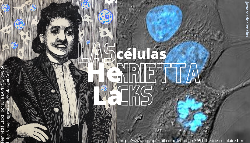
Talking about cell cultures without mentioning Henrietta Lacks is unthinkable, because thousands of scientific and biomedical investigations have been carried out with the cell line that takes its name from the acronym formed with the first two letters of her first name (He) and her last name (La ), but do you know why?

Born in 1920 into a poor African-American family of 10 children who worked in the tobacco plantations of the state of Virginia in the USA, no one imagined that she would become one of the most relevant people in biomedical research… and in a certain way, in “immortal”. This African American, she died in 1951 at the age of 31, a victim of devastating cervical cancer. Without knowing it, it would be the one that would allow, among many other things, to develop vaccines, understand how viruses work or describe a multitude of molecular mechanisms and cellular processes. But how did all this happen?
Before treating the tumor with radium, the surgeon took a sample of Henrietta’s tumor cells without her consent! He gave them to George Otto Gey, a tumor cell culture specialist who worked at the same hospital and had been seeking for years to develop a line of immortal human cells (those capable of dividing indefinitely and remaining alive under culture conditions after a few cell divisions). [5]). Even today, HeLa cells continue to multiply in all cell laboratories around the world.
Henrietta’s were the first immortal human cells to grow in a laboratory and allowed countless experiments to be developed. This represented an enormous advance for medical and biological research. There are currently more than 10,000 patents that include HeLa cells, and it is estimated that scientists have produced more than 20 tons of HeLa cells, even used to investigate human sensitivity to objects as varied as duct tape, glue, cosmetics and many other products.
Although it may seem incredible, it was not until 1996 that the Morehouse School of Medicine in Atlanta and the mayor of Atlanta (USA) belatedly recognized Henrietta Lacks’ family for her posthumous contributions. And it was not until October 2021 when the Director General of the WHO presented a posthumous award to Henrietta’s son, Lawrence Lacks (then 87 years old)—who was accompanied by grandchildren and great-grandchildren—in recognition of the life, legacy and his mother’s contribution to medical science.
The case of HeLa cells is a good example of how cell lines originating from human tissue present bioethical controversies, because they survive the parental organisms and are used to research very lucrative medical treatments after the death of the people who created them. they produced.
APPLICATIONS OF CELL CULTURE
Science routinely uses cell lines to understand the behavior of certain cells, tissues and even entire organisms and their use allows work in a multitude of fields, ranging from basic research to the most applied, both in studies with cells (differentiation, transformation , cell cycle, etc.) as with viruses (component analysis, virus production, etc.)
In BASIC RESEARCH, cell lines allow us to study:
• Intracellular activity, mechanisms involved in different intracellular processes, such as DNA transcription, protein synthesis, energy metabolism, cell cycle, differentiation, apoptosis, etc.
• The intracellular flow of biomolecules, intracellular movements of substances and signals associated with different physiological processes, such as RNA processing, the movement of RNA from the nucleus to the cytoplasm, the movement of proteins to various organelles, assembly and disassembly. of microtubules, etc.
• Genomics and proteomics: genetic analysis, infection, cellular transformation, immortalization, senescence, gene expression, metabolic pathways, etc.
• Cellular ecology: the study of the environmental conditions responsible for the maintenance of cellular functionality and its differentiation, the study of nutritional needs, infections, study of cellular transformation (induced by viruses or chemical agents) or the kinetics of cell population.
• Cellular interactions: embryo induction processes, metabolic cooperation, contact/adhesion inhibition or cell-cell interactions, morphogenesis, cell proliferation, cell invasion.
In APPLIED RESEARCH, cell culture techniques are used in areas as diverse as:
• Virology: Cultivation of animal and plant viruses, production of vaccines, etc.
• Biotechnology: Industrial production of drugs in bioreactors (interferon, insulin, growth hormone, etc.)
• Immunology: Production of monoclonal antibodies, signaling, inflammation phenomena.
• Pharmacology: Effect of various drugs, interactions with the receptor, resistance phenomena, etc.
• Tissue engineering: Production of artificial tissues (skin, cartilage) for the treatment of large burns, grafts or autotransplants, dedifferentiation and induced differentiation, etc.
• Toxicology: Cytotoxicity, mutagenesis, carcinogenesis, etc.
Something very important is that cell culture allows us to minimize the use of research animals and avoid their suffering, because although it cannot always replace the in vivo test, it is a valid alternative in many situations.
Have you been curious and want to know more?
Grades
[1] Royal Decree 664/1997, of May 12, on protection of workers against risks related to exposure to biological agents
[2] The best-known base media for cell culture are Eagle’s Basal Medium, Dulbecco’s Modified Eagle’s Medium (DMEM) and RPMI1640. https://www.studocu.com/es/document/universidad-pablo-de-olavide/cultivos-cellulares/tema-5-laboratorio-de-cultivos-cellulares/6537690
History of cell cultures
[3] Buenas Tareas: Cell Culture https://www.buenastareas.com/ensayos/Cultivo-Celular/55367487.html
[4] EcuRed: Cell Culture https://www.ecured.cu/Cultivo_Celular
[5] Normally, an animal cell can only duplicate itself a certain number of times, because each division shortens the ends of its chromosomes, called “telomeres.” In Henrietta Lacks’ tumor cells there is an enzyme, telomerase, which continually repairs destroyed telomeres, so that the cells can reproduce without limit. https://www.sciencesetavenir.fr/sante/cancer/une-vie-infinie-henrietta-lacks-star-des-labos-malgre-elle_145904
[6] Wikipedia, entry Henrietta Lacks https://es.wikipedia.org/wiki/Henrietta_Lacks
[7] Introduction to cell culture https://www.ehu.eus/biofisica/juanma/mbb/pdf/cultivo_cellular.pdf
[8] SÁNCHEZ, Carlos Manuel. She died of cancer, but her immortal cells have saved millions of people. XLSemanal article https://www.xlsemanal.com/conocer/salud/20181101/celulas-hela-curar-el-cancer-henrietta-lacks.html
Complementary sources
Amanda Capes-Davis, R. Ian Freshney (2021) Freshney’s Culture of Animal Cells: A Manual of Basic Technique and Specialized Applications, 8th Edition, March 2021. Wiley-Blackwell, 832 pp.
Basic aspects of the management and preservation of cell cultures https://www.paho.org/hq/dmdocuments/2010/L%20Brito%20INHRR.pdf
Videos
Cell culture, part I https://www.youtube.com/watch?v=sb0E04_wN7M
Types of cell culture https://www.youtube.com/watch?v=anbcQLJJESo
The Way of All Flesh – Immortal HeLa Cells Documentary https://www.youtube.com/watch?v=cTXaJOk_bjQ&t=4s
