WILD CARD OF CUPS
CONFOCAL MICROSCOPY
Wild Card of Cups: CONFOCAL MICROSCOPY, a very useful technology for neuroscience

Confocal technology is a great advance in optical microscopy, because it solves the problem of observing tissue preparations of a certain thickness, where irregularities make it difficult to find a single plane of focus with sufficient resolution.
Modern biology is based on the study of the internal molecules of the cell and the interactions at the cellular level that allow the construction of multicellular organisms.
Except for some especially large cells—such as plant cells—most are not observable with the naked eye, so to study their structure we need to use amplification systems. To give you an idea, the human eye has a resolution of about 100 microns, which would allow us to see only a few plant cells. Animal cells are 10 times smaller, bacteria are about 100 times smaller, and viruses are nanometers in size, making them completely invisible to the human eye.

Despite these difficulties, human beings have tried to see more than they were capable of: from Confucius, through the reading stones of the Middle Ages or the inventions of Marco Polo, until reaching Galileo Galilei – to whom the discovery of the compound microscope—man has been perfecting the instruments. The first commercial model of compound microscope was put into operation in 1590, dedicated to “cabinets of curiosities” and manufactured by the Jansen brothers, Hans and Zacharias.
By associating two converging lenses in a telescopic tube, the Jansens obtained images magnified up to about 150 times, although not particularly sharp due to the numerous optical aberrations of the system.
The word “microscopy” was first used by members of the Accademia dei Lincei, a scientific society founded in Rome in 1603 by the Italian prince Federico Cesi. Galileo – one of its prominent members – was the one who gave Prince Cesi a microscope with which Federico Stelluti made the observations on the life of bees that he published in his Apiarium (1625).

First microscopist publication: Apiarium, by F. Stelluti (1625)
SOURCE: SOLÍS, C. and SELLÉS, M. (2005) History of Science, p. 338 (Ed. Espasa)
In fact, Hooke is considered the discoverer of the cell due to his work under the microscope, although 200 years had to pass for the establishment of the Cell Theory enunciated by Schleiden, Schwann and Turpin.
And so we could continue describing the slow and continuous advance of optical microscopy from the 15th century to the present day, but today’s entry takes us one step further and brings us CONFOCAL LASER MICROSCOPY.
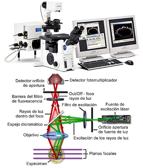
Confocal laser microscopy is a visualization technique for microscopic samples with multiple applications for neuroscience. It works in a similar way to the fluorescence microscope [1], but its main advantage over it is that it allows higher quality images to be obtained through spatial filtering techniques that eliminate light from planes that are out of focus.

Confocal microscopy was patented in 1955 by the American scientist Marvin Minsky in his work on neurons.
Marvin wanted to overcome the limitations of classical photonic (or optical) microscopy, which provides sharper images the thinner the sections of the tissue being examined. When viewing a thick sample under a photon microscope, the image that is in focus is contaminated by the superimposition of out-of-focus tissue elements, both above and below the focused plane, so that the image of the plane that is in focus attempt to observe is deteriorated by blurred or out-of-focus superimposed structures.
Confocal microscopy adds the principle of illuminating the specimen point by point and eliminating light from unfocused planes. This requires a very powerful light source and a filter with a hole (or pinhole) that is placed in the path of the light ray. This allows you to observe both fine optical sections and thicker tissue samples, and even perform 3D reconstructions from serial sections.

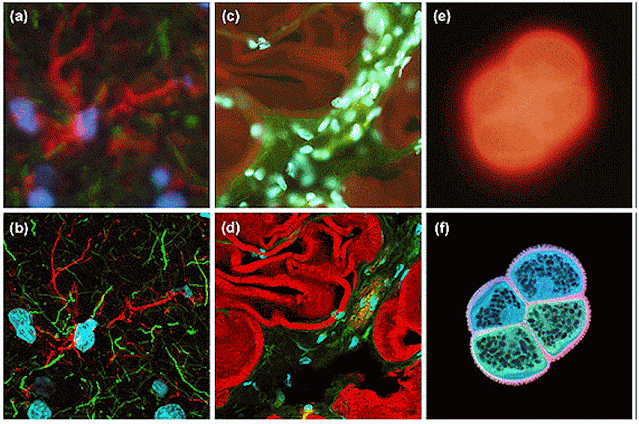
Images taken with a conventional fluorescence microscope (a, c, e) compared to those obtained with a confocal fluorescence microscope (b, d, f) (SOURCE: https://aminoapps.com/c/ciencia/page/blog /microscopy-techniques/q533_W3uRunx41YQ1nNpv5mxJEqJvKRJre)

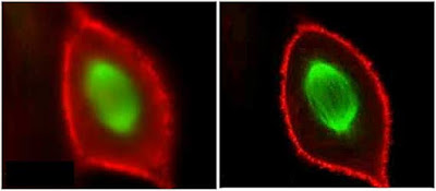
Photograph of a cell in mitosis with double labeling. Chromosomes in green and actin in red.
If you want to know how to work with a confocal microscope, you cannot miss the video that Carmen and Belén (those responsible for the Scientific Imaging and Microscopy Unit of the Cajal Institute, CSIC) have recorded for us. Click on the image to see it:
Have you been curious and want to know more?
[1] Fluorescence is a property of some substances such as collagen, elastin, lignin, chlorophyll and other chemical compounds, which emit light of a defined wavelength after absorbing light of a weaker wavelength. Fluorescence microscopes have xenon or mercury vapor lamps to illuminate the samples. Fluorescence microscopy was discovered by scientists Köhler and Siedentopf in 1908 and is based on the application of a beam of light to a natural substance present in cells or to a fluorescent dye applied to the section of a sample. When carrying out this procedure, it then happens that the sample reflects part of the absorbed energy as light rays, allowing the image of the sample under study to be captured. https://1microscopio.com/de-fluorescencia/#:~:text=Se%20conoce%20como%20microscopio%20de%20fluorescence%2C%20a%20that one, a%20variant%20of%20called%20microscopio%20de%20luz% 20ultraviolet.
