ACE OF CUPS
ASTROCYTES
Ace of cups: Astrocytes, cells that deserve a toast
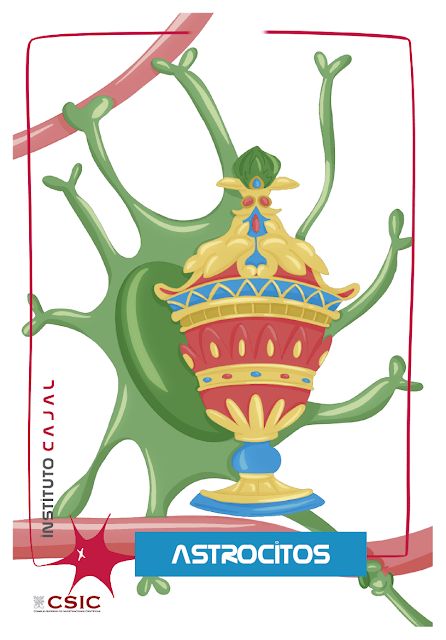
Introducing the Ace of Cups of the neurodeck: the astrocyte. We have chosen astrocytes to represent the suit of cups in our neurodeck because these glial cells support its ability to perform a multitude of essential functions in amazing feet.
Astrocytes are part of a diverse group of cells in the nervous system that, unlike neurons, are not capable of producing action potentials or nerve impulses. They therefore lack axons and dendrites designed to transmit and receive them. This group of cells in nervous tissue are glial cells, also called neuroglia or simply glia.
Glia is present in animals, from the simplest invertebrates to humans. It seems that throughout evolution the glia have been diversifying and specializing until they are a key element in the processes of integrating and processing signals to develop responses that, until very recently, we always believed were exclusive tasks of neurons.
Recent nerve cell counts in the vertebrate brain [1] have confirmed that approximately half of the cells in the mammalian central nervous system are glial cells, which logically suggests that they must play a role in neural circuits. .
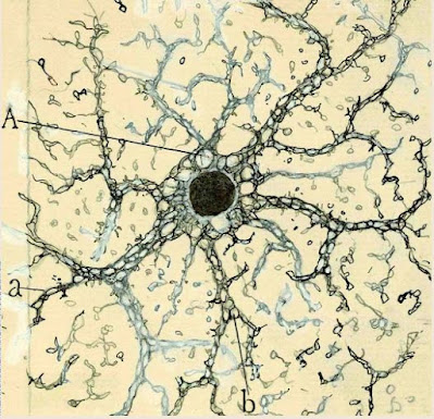
Original drawing by Santiago Ramón y Cajal of a thick neurological cell from the layer of the pyramids of the Horn of Ammon of an adult man
(original image preserved in the Cajal Institute, CSIC (Madrid)
Much less known than neurons, and confined to the central nervous system without direct communication with the outside world, these cells began to intrigue scientists since Golgi techniques for staining tissue samples made them visible under the light microscope, in the middle of the 19th century. XIX century. In 1899 Santiago Ramón y Cajal began to ask himself “What functional significance should we give to neuroglia?” [2], because even then he doubted that they simply played that filler role that had led one of the fathers of cell theory, Rudolph Virchow (1821-1902), to give them the name nervenkitt (“nerve glue” in 1846). German) and later glia (glue in Greek). In his “nutritive theory” of him, Golgi considered that they only gave “logistical support” (food, support structure) to the neurons, but Cajal imagined them with much more prominence (and time has proven him right 😉).
At the end of the 19th century Cajal could only “make more or less rational conjectures”, because the lack of methods left the physiologist “totally disarmed”[2]. However, the development of new tools since the 1980s has allowed neuroscientists today to have an arsenal of techniques to unravel the secrets of neuroglia: the patch-clamp (to record the electrical activity of excitable cells). that produce a small electrical current when stimulated), fluorescent probes (to detect, localize and quantify the dynamics that occur at the cellular scale, thanks to molecules that generate fluorescence signals [3]), new microscopy techniques (confocal, multiphoton…) and, of course, the new techniques of molecular biology and genetic editing.
These scientific-technological advances have shown that Cajal’s intuition was good. Without taking away the morphological and functional prominence of the neuron, today we know that glia orchestrate a series of essential functions for the development and proper functioning of the nervous system, and new roles are increasingly being discovered in pathological situations or even related to processing. and the integration of information that neurons make. Divided into two types, microglia (of non-nervous origin) and macroglia (with the same embryonic origin as neurons), glial cells are no longer extraneous and are now as important players in the functioning of the nervous system as neurons.
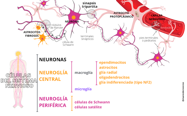
Santiago Ramón y Cajal imagined the first technique to specifically stain astrocytes [4] thanks to the gliofibrillary acid protein (GFAP) that forms the cytoskeleton responsible for maintaining the structure of these cells and that forms the glial fibrils in the cells. extensions of the cytoplasm. Currently, GFAP allows the identification of astrocytes using immunohistochemical markers.
The number of astrocytes varies depending on the area of the nervous system in which they are found (from 20% to 85%), but in the brain and spinal cord of mammals they represent more than 50% of the neuroglia [5] ,[6]. They are the largest glial cells (diameter of their cell body between 18 and 20 micrometers).
Although they are generally recognized by the presence of a large oval or lobed nucleus and the star-shaped shape that inspired their name, the shape and appearance of astrocytes can vary depending on where they are found. There are highly modified astrocytes in the cerebellum (Bergmann glia), in the retina (Müller glia) and their appearance is different in the gray and white matter of the brain, as Andriezen demonstrated in 1893 [7].
In the gray matter, protoplasmic astrocytes are abundant, with short and highly branched processes surrounding neuronal somata and dendrites in tripartite synapses. Fibrous astrocytes are associated with neuronal axons in the white matter and have much fewer and longer extensions, almost unbranched, in addition to a cytoplasm full of filaments. It seems that there is a third type of reactive astrocytes that, in pathological situations, would be capable of assuming defensive functions together with microglia to deal with certain brain injuries, forming the so-called glial scar.
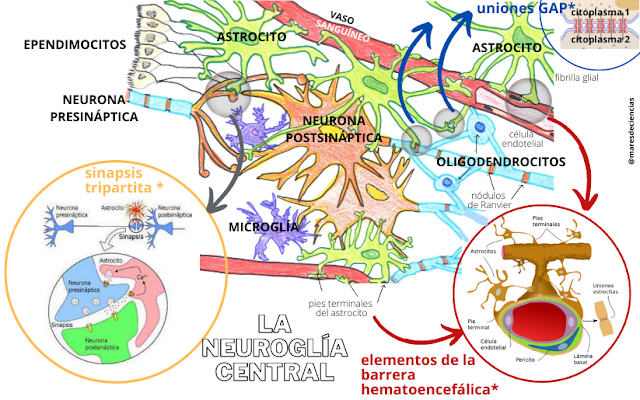
Another characteristic of astrocytes is that they are cells capable of forming extensive networks where they keep their cytoplasms interconnected thanks to GAP junctions (small tunnels in the plasma membrane formed by connexin protein that allow ions and small molecules to pass in both directions) [ 9].
To communicate, astrocytes also produce and release chemical messengers (called gliotransmitters) that allow a population of astrocytes to act in a synchronized and coordinated manner to participate with neurons in the processing of information.
Among other researchers at the Cajal Institute, Alfonso Araque and his team study the modalities of the “conversation” established in what they have called “tripartite synapses”, where astrocytes “comment” and “moderate” the dialogue between neurons, and They investigate its implications in the development of diseases of the nervous system. They have established that astrocytes have a form of excitability based on variations in the internal concentration of calcium (Ca2+), so that they communicate with each other through intercellular calcium waves. As astrocytes also have receptors for neurotransmitters produced by neurons in their membrane, the nervous impulse can excite them when they envelope the synapse and cause them to release gliotransmitters (mainly ATP and glutamate), which in turn will modulate neuronal synapses.
It has been observed that neurons not only release chemical messengers (neurotransmitters) at synapses, but also outside them, suggesting that the functions of neuron-glia communication are much more complex than simple collaboration in synaptic transmission. It appears that glia can regulate synapse formation, control synaptic strength, and participate in information processing by coordinating activity between different groups of neurons. And conversely, nerve impulse activity can regulate a wide range of glial activities, including proliferation, differentiation and myelination [2],[5], [10].
With so many capabilities, the “catalog of services” that astrocytes offer us is impressive [12] [13]:
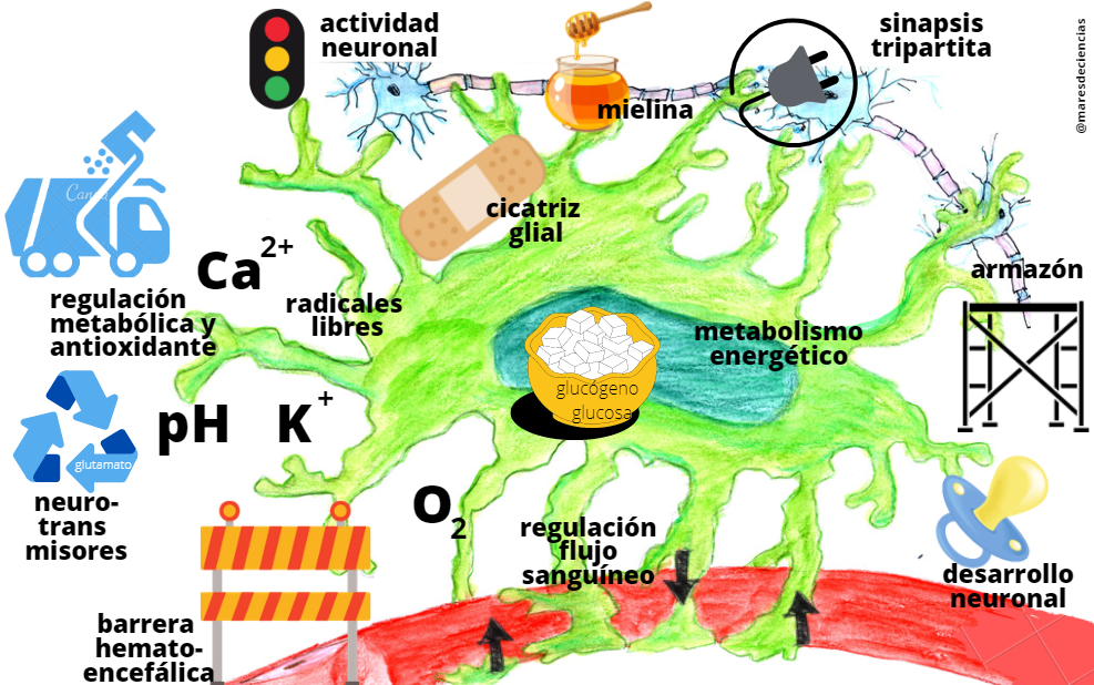
1. STRUCTURAL FRAME. Astrocytes participate in the physical structuring of the brain and play a secondary mechanical support role.
2. PROTECTION. The endfeet of astrocytes form a continuous layer that surrounds the blood vessels at the periphery of the central nervous system, thereby contributing to the blood-brain barrier that separates blood from the extracellular brain fluid in the central nervous system. Here astrocytes prevent the passage of substances potentially dangerous to the brain and allow access to others, such as water, oxygen or small fat-soluble substances. Recently, the glymphatic system has been discovered, a brain drainage pathway that depends on the proper functioning of neuroglia and especially astrocytes.
3. METABOLIC REGULATION AND ANTIOXIDANT ACTION. Astrocytes are responsible for controlling the amount of substances present in the environment of neurons so that they function correctly and generate clean signals. Specifically, they control pH, potassium (K+) and calcium (Ca2+) levels, capture free radicals (thanks to glutathione), and intervene in the recycling metabolism of neurotransmitters (for example, toxic excess glutamate is absorbed). incorporates glutamate/glutamine into the cycle).
4. ENERGY METABOLISM. Astrocytes manage their cytoplasmic glycogen reserve to maintain it at levels that allow them to cope with the demand for glucose in periods of high neuronal activity. They can also be responsible for transforming glucose into lactate available to the neuron (for the Krebs cycle).
5. CONTROL AND REGULATION OF BLOOD FLOW that reaches the brain and neurons to provide them with the necessary oxygen (O2). The local increase in blood flow supplies more oxygen and nutrients to active brain regions that need more energy.
6. MODULATION OF NEURONAL ACTIVITY. Astrocytes regulate the formation, maturation, maintenance and stability of synapses so that they function correctly, thereby maintaining the connectivity of neuronal circuits, such as those associated with learning and memory. Thus, astrocytes intervene in how the nervous information handled by neurons is processed.
7. REGULATION OF SYNAPSES. In gray matter, astrocytes are closely associated with neuronal membranes, especially in synaptic areas. This is where the terminal feet completely or partially envelop the presynaptic terminals and postsynaptic structures, forming the tripartite synapse.
8. NEURONAL DEVELOPMENT AND PLASTICITY. Astrocytes play an important role in the embryonic development of nervous tissue, and are responsible, for example, for guiding axonal growth or helping neurons and other nerve cells reach the places in the cerebral cortex where they should be.
9. COORDINATION OF MYELIN PRODUCTION. Astrocytes are responsible for transforming adenosine triphosphate (ATP) [11] that releases the electrical activity of neurons into a protein that stimulates the production of myelin in oligodendrocytes.
10. DEFENSE. Astrocytes are involved in inflammatory processes of immunological reactivity of the brain. In response to brain injuries and thanks to a certain capacity to generate an immune response (astrocytes are immunocompetent), they participate with microglia in the formation of the glial scar.
Incredible true? You’re probably thinking that these astrocytes are extraordinary nerve cells that deserve a toast… with the best glasses in the neurodeck!
Have you been curious and want to know more?
GRADES
[1] VON BARTHELD, Christopher S., Jami BAHNEY, Suzana HERCULANO-HOUZEL (2016). The search for true numbers of neurons and glial cells in the human brain: A review of 150 years of cell counting. The Journal of Comparative Neurology, vol. 524 (18), pp. 3865-3895. https://onlinelibrary.wiley.com/doi/10.1002/cne.24040
[2] Cajal proposed the “isolation theory”, in which he imagined astrocytes as insulating elements for different neurons. ARAQUE, Alfonso and Marta NAVARRETE (2013). The yesterday and today of astrocytes. Mind and Brain No. 59, pp. 18-23. https://www.investigacionyciencia.es/revistas/mente-y-cerebro/evolucin-del-pensamiento-575/el-ayer-y-hoy-de-los-astrocitos-11083
[3] Calcium imaging techniques are especially important in neuroscience. In nervous tissue, calcium (Ca2+) is a very important ion that is interesting to detect. Fluorescence-based methods are selective, very sensitive, fast and affordable. Chemosensors are molecular receptors that bind only to the molecule or ion we are interested in studying and, in doing so, produce some type of measurable signal, such as fluorescence, redox potential or absorption spectra. In a chemosensor or fluorescent probe there is a receptor responsible for recognition and a fluorophore that signals this recognition with changes in its fluorescence.
[4] This is the gold chloride sublimation technique. For a review of the different methods of staining nerve cells, see: Brain Imaging Techniques, by José Méndez Venegas (Faculty of Psychology, UNAM, Mexico).
[5] CALDERÓN ÁLVAREZ-TOSTADO, J.L. and RIVERA SILLA, G. (2012). The astrocyte: a star behind the scenes. Science, January-March 2012, pp. 68-74. https://www.revistaciencia.amc.edu.mx/images/revista/63_1/PDF/10_698_Astrocito.pdf#:~:text=El%20astrocito%3A%20una%20estrella%20tras%20bambalinas%20central%20es,know %20in detail%20your%20behavior.%20%C2%BFBlanco%20for%20treatments%20m%C3%A9medical%3F
[6] MEGÍAS M., MOLIST P., POMBAL M. A. (2019). Atlas of plant and animal histology. Cell types. Astrocyte. http://mmegias.webs.uvigo.es/8-tipos-cellulares/astrocito.php
[7] Cajal cites in his work “Texture of the nervous system of man and vertebrates” (1899 – 1904) the works of Andriezen, who in 1893 had distinguished a fibrous glia (in German Langstrahler or “long projectors”) that It is located in the white matter of the brain from a protoplasmic glia (in German Kurzstrahler, or “short projectors”) in the gray matter. In MARTÍNEZ-TAPIA, R. J. et al. (2018) A new brain drainage pathway: the glymphatic system. Historical and conceptual review Mexican Journal of Neuroscience January-February, 2018; 19(1), pp. 104-116. https://www.medigraphic.com/pdfs/revmexneu/rmn-2018/rmn181j.pdf
[8] MEDEROS CRESPO, Sara (2019). Astrocyte-interneuron communication and information processing in neuronal networks. Doctoral Thesis directed by Dr. Gertrudis PEREA PARRILLA, from the Cajal Institute (CSIC). Complutense University of Madrid. Faculty of Biological Sciences, Department of Biochemistry and Molecular Biology. 198 pp. https://digital.csic.es/handle/10261/210592
[9] We already saw in the entry of the Ace of Pentacles that this type of GAP junctions are what allow the very rapid and two-way circulation of the nervous impulse in the electrical synapses.
[10] FIELDS, R. D. & B. STEVENS-GRAHAM (2002). New Insights into Neuron-Glia Communication. Science (298), pp. 556-562 https://www.science.org/doi/10.1126/science.298.5593.556
[11] What is ATP? (cienciaybiologia.com) https://cienciaybiologia.com/que-es-el-atp/
[12] YANGÜAS CASÁS, Natalia (2015). Effect of taurorsodeoxycholate on the regulation of acute neuroinflammation. Doctoral Thesis directed by Dr. Lorenzo Romero Ramírez and Prof. Manuel Nieto Sampedro (CSIC). Autonomous University of Madrid, Faculty of Medicine, Department of Biochemistry. https://repositorio.uam.es/handle/10486/668514?show=full
[13] YANGUAS CASÁS, Natalia (2020). Neuroscience for dummies. Eleventh night of European researchers, Madrid. 2020. Cajal Institute YouTube Channel
https://youtu.be/1SZ2HVnGwIk?list=PLEU_4DYTRsCHPRnT2mlufUmpb_J0KlwmY&t=1311
SOURCES
COVELO FERNÁNDEZ, Ana (2015). Short- and long-term synaptic modulation mediated by astrocytes in the hippocampus. Doctoral Thesis directed by Dr. Alfonso ARAQUE ALMENDROS, from the Cajal Institute (CSIC). Autonomous University of Madrid. Faculty of Medicine, Department of Anatomy, Histology and Neuroscience. 109 pp. https://repositorio.uam.es/bitstream/handle/10486/669246/covelo_fernandez_ana.pdf?sequence=1
JÄKEL, Sarah and Leda DIMOU (2017). Glial Cells and Their Function in the Adult Brain: A Journey through the History of Their Ablation. Frontiers in Cellular Neuroscience. February 2017, vol. 11, no. 24. https://doi.org/10.3389/fncel.2017.00024
LEBRERO CIA, Carmen (2017), Modulation of intercellular communication mediated by GAP junctions in the HEK 293 cell line. Final Degree Project in Biotechnology, Miguel Hernández University of Elche, 35 pp. http://dspace.umh.es/bitstream/11000/4145/1/TFG%20Lebrero%20Cia%20Carmen.pdf
PÉREZ CAPOTE, Kamil (2006). Response of glial cells to neuronal damage in vitro. Introduction (55 pp). Doctoral Thesis. University of Barcelona. https://digital.csic.es/bitstream/10261/91949/4/1_INTRODUCCION.pdf
REYES-HARO, Daniel, Larissa BULAVINA and Tatyana PIVNEVA (2014). Glia, the glue of ideas. Science, April June 2014, pp. 12-18.
Videos:
Synapsis EMP YouTube Channel. Astroglia. https://www.youtube.com/watch?v=crFp-Hja9i4
UnProfesor YouTube Channel What is the blood-brain barrier? https://www.unprofesor.com/ciencias-naturales/que-es-la-barrera-hematoencefalica-2394.html
Websites:
https://celulasgliales.com/
https://w3.ual.es/personal/pnieto/Neurona%20y%20glia/macroglia.htm
