ACE OF CLUBS
THE OLIGODENDROCYTE
Ace of clubs: oligodendrocytes, myelin producers

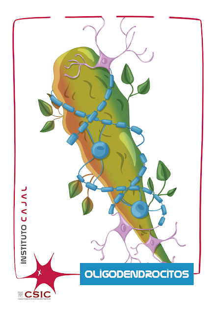
We present the Ace of clubs of the neurodeck: the oligodendrocyte, a highly coiled glial cell, a specialist in covering neuronal axons with insulating and protective myelin that, when missing, paints wands.
In the Neuroglial Networks Laboratory of the Cajal Institute, Sara Mederos Crespo reminds us that already in 1909 Ramón y Cajal in his “Histologie du systeme nerveux de l’homme et des vertébrés” named the glia cells and gave them a much more important role. prominent in brain function than those of filling and trophic support initially attributed by Rudolph Virchow in 1846 [1].
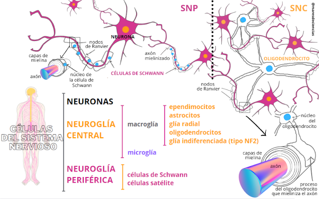
Glial cells encompass several cell types that can be differentiated into two groups: microglia and macroglia. Microglia are mainly responsible for the immunological reaction within the central nervous system (and we will dedicate the Ace of Swords entry to them), while the varied group of macroglia cells (which includes astrocytes, ependymocytes, oligodendrocytes and Schwann cells) It is responsible for various functions.

While an oligodendrocyte can extend its processes (processes) to wrap myelin up to 50 axons, in the nerves of the peripheral nervous system the axons are covered by a succession of Schwann cells, in which one cell surrounds a single axon.

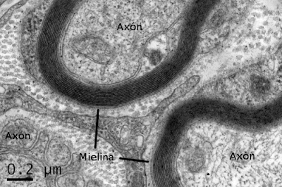
Myelin sheath in a peripheral nerve seen under an electron microscope
Source: https://mmegias.webs.uvigo.es/5-celulas/3-membrana_cellular.php
Oligodendrocytes are characterized by:
- Being particularly abundant in the nervous centers (for example, they are very abundant in the corpus callosum), mainly forming rows parallel to the nerve fibers in the white matter, and groupings around the neurons in the gray matter.
- Have a round, voluminous and dense core.
Present few branches of the cytoplasm, generally fine and with few granulations (gliosomes) inside the cytoplasm.
Wrap around the nerve fibers of the white matter. - Due to their location and characteristics, oligodendrocytes can be of three types [2]:
- Satellite oligodenrocytes, very small (10 µm) and only present in the gray matter. As they surround the body of the neuron, it is assumed that they participate in proper neuronal functioning, but many things about them are still unknown.
- Interfascicular oligodendrocytes, somewhat larger in size (20 µm) and with a very voluminous nucleus in adults. They are located in the white matter, where they run parallel along the axons that myelinate and maintain myelination.
- Intermediate oligodendrocytes are found in both gray and white matter. It is thought that these may be precursor cells of interfascicular and satellite oligodendrocytes.
In addition to producing and maintaining myelin sheaths and regulating the function of axons, it seems that they would also be involved in the control of the ionic environment, referring above all to the transport of water and chlorine [2] and the regulation of the expression of the channels. sodium and potassium.
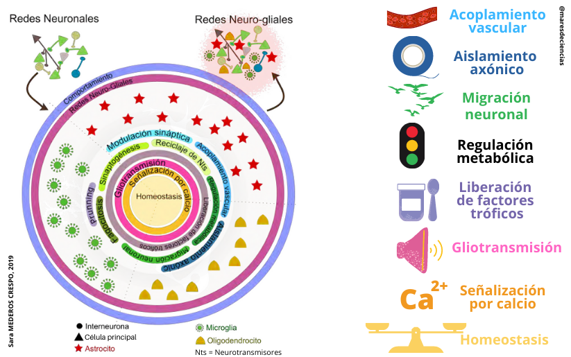
From the metabolic point of view, let us remember that astrocytes nourish the neuron, synthesize and recycle neurotransmitters and participate in the repair and modulation of neuronal and synaptic activity. Oligodendrocytes generate the myelin sheath, contribute to the signaling and growth of neurons, and collaborate in their nutrition and maintenance. All these functions are summarized in this diagram [1]:

LEFT: Neuro-glial networks: signaling units in the brain [1]. RIGHT: Functions of oligodendrocytes, which generate the myelin sheath, contribute to signaling and neuron growth.
For its part, microglia play an important role in the immune response of the CNS. https://digital.csic.es/handle/10261/210592
Pío del Río-Hortega was always interested in the Golgi metallic impregnation techniques improved by Cajal (with uranium nitrate or sublimated gold chloride) and Achúcarro (based on tannin and ammoniacal silver). He created four variants of Achúcarro’s technique to identify the protoplasmic and fibrous astrocytes that were the “second element” that accompanied neurons (or “first element”). However, these histological methods only partially stained the rest of the glial cells of the central nervous system, which are very abundant in the white matter of the brain, and which under the microscope were seen as small, unbranched (adendritic) cells. Río-Hortega continued to investigate new staining techniques to find out the true nature of what Ramón y Cajal had called the “third element”, until he developed the ammoniacal silver carbonate method. He was thus able to demonstrate that the third Cajalian element corresponded to 2 cell types: microglia (or Hortega cells) and oligodendroglia [3] [4] [5].
Starting in 1921, the silver carbonate method and a variant of the Golgi method without osmium allowed Río-Hortega to study those poorly branched but very abundant cells in the white matter that he initially called “interfascicular glia” and later oligodendroglia. .
Río-Hortega was a pioneer in the knowledge of oligodendrocytes and, despite the great variability of the results he obtained with his staining methods, he intuited their relationship with the myelin coating of the axons. In 1922 he pointed out that the function of oligodendrocytes in the central nervous system was similar to that of Schwann cells in the peripheral nervous system: to produce the myelin sheath that protects and insulates the transmitting elements of neurons – axons -, although the demonstration had to wait for the development of electron microscopy in the 1960s [6].
Different immunocytochemical techniques are currently used to identify oligodendrocytes thanks to a range of antibodies against surface and intracellular markers, and to the techniques of electron microscopy, immunomicroscopy and three-dimensional electron tomography [6][7], because oligodendrocytes still keep many secrets. In the field of demyelinating diseases, fundamental research is the basis on which therapeutic progress aimed at both preventing demyelination and promoting remyelination must be based.
Having a limited regenerative capacity, oligodendrocytes are the most vulnerable cell type and the first nervous elements to degenerate in diseases of the central nervous system. In the adult brain of a human being, there are between 5% and 8% of cells called oligodendrocyte precursor cells (OPCs), capable of producing immature oligodendrocytes that migrate following very specific and chemically orchestrated routes to the brain. place where mature myelinating oligodendrocytes become, capable of remyelinating the injured area. A remyelination rate of 12% is estimated to achieve significant improvement in symptoms due to demyelination. In healthy organisms, this regenerative capacity is sufficient, but not in a disease situation. For this reason, at the Cajal Institute, cutting-edge lines of research are pursued that seek to identify cellular signals that can be exploited, for example through drugs that cause a type of cell to evolve towards the type of cell capable of promoting remyelination [9]. [10].
Have you been curious and want to know more?
SOURCES CITED
[1] MEDEROS CRESPO, Sara (2019). Astrocyte-interneuron communication and information processing in neuronal networks. Doctoral Thesis directed by Dr. Gertrudis PEREA PARRILLA, from the Cajal Institute (CSIC). Complutense University of Madrid. Faculty of Biological Sciences, Department of Biochemistry and Molecular Biology. 198 pp. https://digital.csic.es/handle/10261/210592
[2] PÉREZ CAPOTE, Kamil (2006). Response of glial cells to neuronal damage in vitro. Introduction (55 pp). Doctoral Thesis. University of Barcelona. https://digital.csic.es/bitstream/10261/91949/4/1_INTRODUCCION.pdf
[3] SIERRA, Amanda, PAOLICELLI Rosa C., and KETTENMANN H. (2019). One Hundred Years of Microglia: Milestones in a Century of Microglial Research. Trends in Neurosciences, November, Vol. 42, No. 11, pp. 778-792. https://doi.org/10.1016/j.tins.2019.09.004
[4 ] DEL RÍO-HORTEGA, Pío (1919). Rapid staining of normal and pathological tissues with ammoniacal silver carbonate. Works of the Histopathology Laboratory of the Board for the Expansion of Studies No. 7. Bulletin of the Spanish Society of Biology, Vol. . 235-243
https://arbor.revistas.csic.es/index.php/arbor/article/view/432/434
[5] DEL RÍO-HORTEGA, Pío (1924). What should be understood by the Third Element of the nervous centers. Bulletin of the Spanish Society of Biology, Vol. 245-248
https://arbor.revistas.csic.es/index.php/arbor/article/view/433/435
[6] FARIÑA GONZÁLEZ, Juliana and ESCALONA ZAPATA, J. (2010). The work of Pío del Río-Hortega and its consequences in neuropathology https://pio-del-rio-hortega.blogspot.com/
[7] DOMÍNGUEZ, Raúl O. (2016). Prof. Pío del Río-Hortega: from oligodendroglia to demyelination-remyelination. Previous history and exile in Argentina 1940-1945. Argentine Neurology, Vol. 8. No. 1, pp. 61-64 (January-March) http://dx.doi.org/10.1016/j.neuarg.2015.04.002
[8] PÉREZ CERDÁ, Fernando, Mª Victoria SÁNCHEZ GÓMEZ and Carlos MATUTE (2015). Pío del Río-Hortega and the discovery of the oligodendrocytes. Frontiers in Neuroanatomy, Volume 9 (July 2015), Article 92 https://doi.org/10.3389/fnana.2015.00092
[9] DE CASTRO SOUBRIET, Fernando. Oligodendrocyte precursors (EM online, May 2016) https://www.youtube.com/watch?v=7X_RxZGW9wk&t=35s
[10] Remyelinating therapies Canal Sinapis EMP – YouTube https://www.youtube.com/watch?v=oJKLripB9Os
OTHER LINKS
Cajal Institute YouTube Channel
NEUROSCIENCE FOR DUMMIES, with Natalia Yangüas Casás.
https://youtu.be/1SZ2HVnGwIk?list=PLEU_4DYTRsCHPRnT2mlufUmpb_J0KlwmY&t=1541
The brain dictionary. YouTube Channel Cerebrotes, by Clara García
Myelin https://www.youtube.com/watch?v=HswTL7F0u0I&t=330s
Glial Cells Blog https://celulasgliales.com/oligodendrocitos/
JÄKEL, Sarah and Leda DIMOU (2017). Glial Cells and Their Function in the Adult Brain: A Journey through the History of Their Ablation. Frontiers in Cellular Neuroscience. February 2017, vol. 11, no. 24. https://doi.org/10.3389/fncel.2017.00024
DO YOU WANT TO CHECK WHAT YOU KNOW ABOUT ASTROCYTES AND OLIGODENDROCYTES? Well, here are a series of questions. Luck!
